
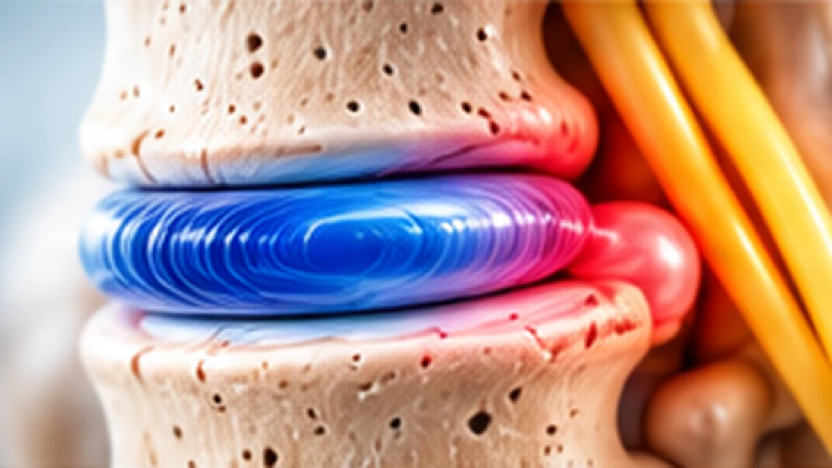
An L4–L5 disc finding can sound alarming, particularly when an MRI report uses terms such as bulge, protrusion, extrusion or nerve-root contact. These words describe anatomy, not a complete diagnosis. The right plan depends on the symptoms, examination, daily limitations, health history and whether the scan finding matches what the clinician observes.
Key points
- An MRI phrase alone does not decide treatment or prove that the disc is causing pain.
- Many patients can begin with non-surgical care and a guided return to activity.
- Persistent disabling nerve pain or progressive neurological changes warrant specialist assessment.
- New bladder or bowel problems, saddle numbness or rapidly worsening weakness require emergency care.

What does L4–L5 mean?
The lumbar spine is the lower part of the back. L4 and L5 are the fourth and fifth lumbar vertebrae, and the disc between them helps distribute load and allows movement. A disc has a tougher outer ring and a softer centre. With age, repeated loading, injury or a combination of factors, the disc may lose water content and change shape. A bulge generally describes a broader extension of disc tissue beyond its usual boundary; a herniation usually describes more localised displacement. Reports may use additional terms to describe the shape and direction. Those labels are useful, but they do not independently tell us how much pain a person should feel. Many adults have disc changes without symptoms. Conversely, a relatively small finding can matter if it irritates a sensitive nerve root. The clinician therefore needs to match the scan to the history and examination rather than treating the report as a verdict.
Symptoms and patterns that may occur
An L4–L5 problem may cause lower-back pain, but symptoms can also travel into the buttock, thigh, shin or foot when a nerve root is irritated. People describe burning, electric-shock sensations, pins and needles, numbness or pain that worsens with coughing, sneezing, bending or prolonged sitting. Some notice weakness when lifting the foot or big toe, although the pattern varies and other conditions can produce similar symptoms. Pain limited to the back does not automatically mean a disc herniation, and leg pain does not automatically prove one. Hip problems, peripheral nerve disorders and other spinal conditions can overlap. Tell the clinician where symptoms begin and travel, whether they are constant or intermittent, and what changes them. Report new weakness rather than simply describing pain intensity. A clear symptom map can be more useful than trying to interpret medical terminology alone.
Why the same MRI finding affects people differently
Two people can have similar MRI wording but very different experiences. A nerve may be compressed, inflamed or not meaningfully affected at all. The nervous system’s sensitivity, general health, sleep, stress, activity and previous episodes can influence how symptoms behave. This does not make pain imaginary. It means pain is a biological experience shaped by more than a single image. The location and direction of a disc change also matter, as does the amount of space available around the nerve. Clinicians assess function: walking distance, ability to sit, sleep quality, work tasks and strength. They also look for signs that symptoms may arise from another source. A treatment plan should address the person, not only the scan. Be cautious of claims that a particular MRI phrase inevitably leads to paralysis or surgery. Such predictions cannot be made from the phrase alone.
How a specialist assesses an L4–L5 problem
Assessment usually starts with the timeline: when pain began, whether there was an injury, how symptoms have changed and what treatments have already been tried. The clinician asks about walking, sleep, work, medication, medical conditions and previous spine problems. Examination may include gait, lower-limb strength, sensation, reflexes and movements that reproduce or ease symptoms. Hip and peripheral nerve assessment may be needed because not every leg symptom comes from the spine. Bring the full MRI report and, when available, the images themselves. A previous scan may help show whether a finding is longstanding or new. Tell the clinician about fever, recent infection, cancer history, unexplained weight loss, significant trauma or immune-suppressing medicines. These details may change the urgency or type of investigation. The goal is to establish a working diagnosis and identify any warning signs before discussing treatment options.
What an MRI can and cannot tell you
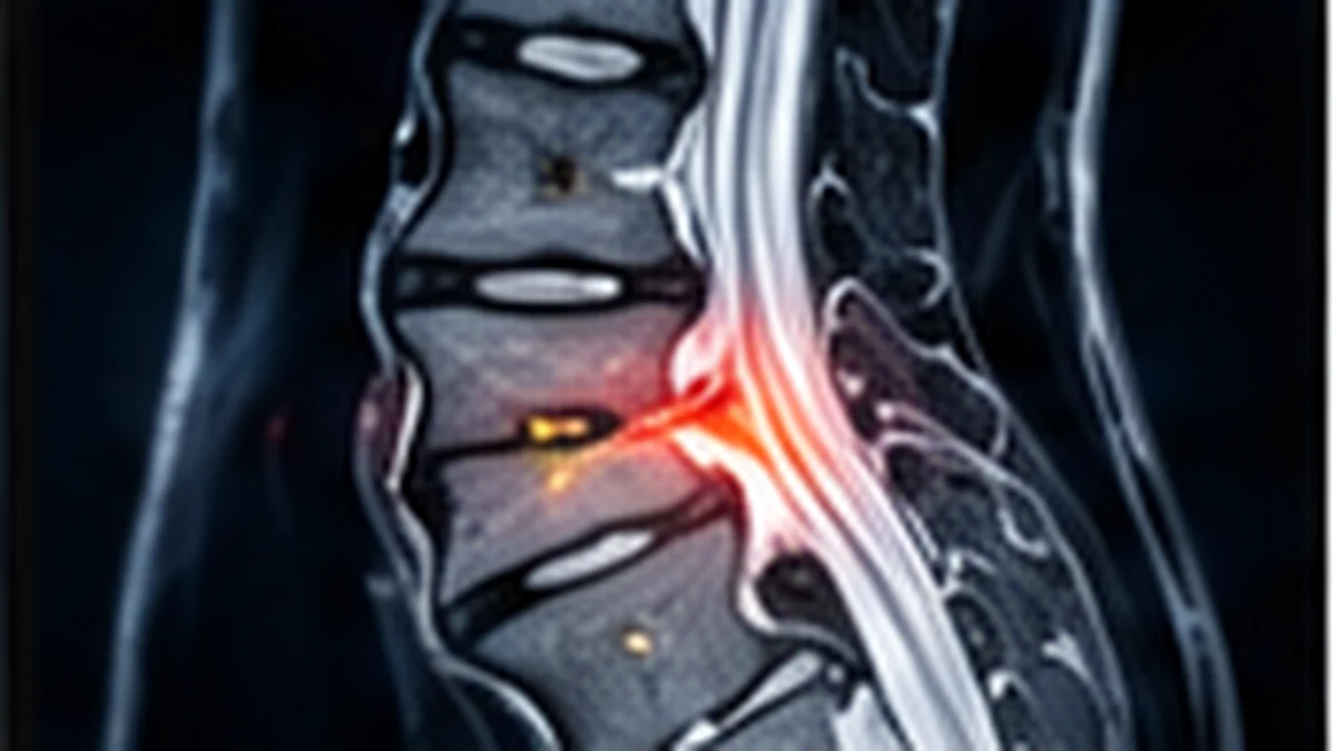
MRI can show discs, nerve roots, the spinal canal and many soft-tissue structures. It can help identify whether a disc protrusion appears to contact or compress a nerve. Yet MRI is a static picture taken at one point in time. It does not directly measure pain, and it cannot reliably predict the exact recovery date. Age-related disc changes are common in people who have no back pain. A report may list several abnormalities, but only some may be relevant to the current symptoms. The most useful question is not simply “Is the scan abnormal?” but “Does this finding fit the symptoms and examination, and would it change management?” Imaging is generally considered when the result is likely to affect decisions, when symptoms persist despite appropriate care, or when urgent warning signs require investigation. Avoid repeating scans solely to track pain unless a clinician explains why a new study is needed.
Non-surgical treatment and early recovery
When there are no emergency features or progressive major neurological deficits, clinicians often begin with non-surgical treatment. This may include explanation of the condition, activity modification, suitable pain relief prescribed after considering individual risks, and physiotherapy. The objective is to keep you safely moving while symptoms settle and capacity improves. Short periods of rest may help during a flare, but prolonged bed rest can lead to stiffness, reduced strength and fear of movement. Walking for manageable intervals, changing positions regularly and gradually returning to ordinary tasks may be more useful than waiting for all pain to disappear. There is no single exercise routine suitable for everyone. Movements that sharply increase radiating pain, create new numbness or worsen weakness should be discussed with the treating clinician. People with kidney disease, stomach ulcers, blood-thinning medication, pregnancy or other conditions should not self-select pain medicines without professional advice.
The role of physiotherapy and home activity
A physiotherapy programme should be matched to the current stage of symptoms, examination and goals. Early sessions may focus on comfortable movement, walking tolerance, positions that ease symptoms and safe ways to manage daily tasks. As symptoms permit, the plan may progress to trunk and hip strength, mobility, balance and conditioning. The aim is not to force the spine into a supposedly perfect posture; it is to build confidence and capacity for normal life. A home programme works best when it is realistic and performed consistently rather than aggressively. Tell the therapist if pain travels farther down the leg, weakness appears or symptoms remain substantially worse after an exercise. Workstation changes, lifting technique and breaks from prolonged sitting may reduce repeated aggravation, but they cannot guarantee that a disc will return to a particular shape. Progress is better judged by function and symptom trend than by one difficult day.
Medicines and injection options
Medication may help some people participate in sleep, walking and rehabilitation, but it does not suit everyone and does not remove the need for assessment. A clinician may consider short-term options based on the suspected cause of pain, other medical conditions and possible interactions. Do not combine medicines or increase doses because a friend had a different prescription. Some medicines have important kidney, stomach, blood pressure, sedation or bleeding risks. In selected cases, an image-guided injection may be discussed for severe nerve-related pain. Benefits can vary, and an injection is not a permanent repair of the disc. The clinician should explain the intended target, expected duration of benefit, risks, alternatives and what the next step would be if it does not help. Ask whether the goal is short-term symptom control to support rehabilitation or another specific clinical objective. Procedures should be selected for a defined reason, not simply because an MRI looks dramatic.
When surgery may be considered
Surgery may be discussed when disabling leg pain continues despite a reasonable trial of suitable non-surgical care, examination and imaging identify a matching compression, or a neurological deficit changes the balance of risks and benefits. The decision depends on symptom severity, duration, functional impact, general health and personal goals. Different procedures address different problems, so it is important to understand the proposed operation rather than assuming all “disc surgeries” are the same. A surgeon should explain which symptoms are most likely to improve, which may persist, the expected recovery process, possible complications and non-operative alternatives. Surgery can be effective for appropriately selected nerve compression, but it is not a guarantee of a completely pain-free back. Back pain, leg pain, numbness and weakness may recover at different rates. Urgent neurological symptoms require a different pathway from a planned decision after weeks of symptoms. A second opinion can be reasonable when the diagnosis or recommendation remains unclear.
Warning signs that need urgent attention
Seek emergency medical assessment if back or leg symptoms are accompanied by new difficulty passing urine, loss of bladder or bowel control, numbness around the groin or saddle area, or rapidly worsening leg weakness. These can be signs of serious nerve compression and should not wait for a routine appointment or an online response. Prompt assessment is also important after major trauma or when severe pain occurs with fever, recent serious infection, a history of cancer, unexplained weight loss or other concerning systemic symptoms. These features do not all mean the same diagnosis, but they may require urgent investigation. Do not drive yourself if weakness or pain makes driving unsafe. If symptoms are changing quickly, tell the emergency team exactly when the change began. Early assessment matters because some causes of nerve dysfunction are time-sensitive.
Returning to work, sleep and everyday life
Recovery is rarely a perfectly straight line. A better morning may be followed by a difficult afternoon, particularly when activity increases. That does not automatically mean new damage has occurred. Increase walking, household tasks and work demands in manageable steps, and plan breaks before symptoms become overwhelming. For desk work, alternate positions and take brief movement breaks rather than searching for one magic chair. When lifting is unavoidable, keep the load close, avoid sudden twisting under heavy load and ask for help with awkward objects during a flare. For sleep, choose a position that is comfortable; some people prefer side-lying with a pillow between the knees, while others feel better on their back with support beneath the knees. There is no universally correct posture. Track useful measures such as walking time, sleep and ability to complete tasks. These can help the clinician judge progress more accurately than pain scores alone.
Preparing for a spine consultation in Kharghar or Navi Mumbai
For a consultation about L4–L5 disc bulge treatment in Kharghar or Navi Mumbai, bring the MRI report and images, a list of medicines, previous treatment details and notes about symptom changes. Write down what you can no longer do comfortably, how far you can walk and whether symptoms affect sleep or work. Mention any new numbness or weakness clearly. Useful questions include: What diagnosis best explains my symptoms? Does the MRI match the examination? What should I try first, and how will we measure progress? Which symptoms should prompt urgent review? If surgery is being considered, what specific problem would it address and what are the alternatives? The clinic team can explain options after assessing your individual situation. A useful consultation should leave you with a clear plan, realistic expectations and instructions for when to seek help, rather than simply a label from a scan report.
Frequently asked questions
Many patients improve with time and appropriate non-surgical care. Whether surgery is relevant depends on symptoms, examination, imaging and neurological changes.
No. MRI findings are common in people without symptoms. The scan must match the clinical picture and change management before it guides a treatment decision.
There is no single timeline. Recovery depends on the cause, severity, treatment, general health and the demands of daily life. A clinician can help set meaningful milestones.
Gentle activity is often encouraged when appropriate, but the amount and pace should fit symptoms and clinical advice. New weakness or rapidly worsening symptoms require assessment.
New bladder or bowel dysfunction, saddle numbness or rapidly worsening weakness requires emergency assessment.
Questions about your spine symptoms?
Arrange a consultation to discuss symptoms, examination findings and available treatment options with the clinic team.
Educational information only; this page does not replace an individual medical assessment. Medical information is provided for general education and does not replace an individual clinical assessment.