
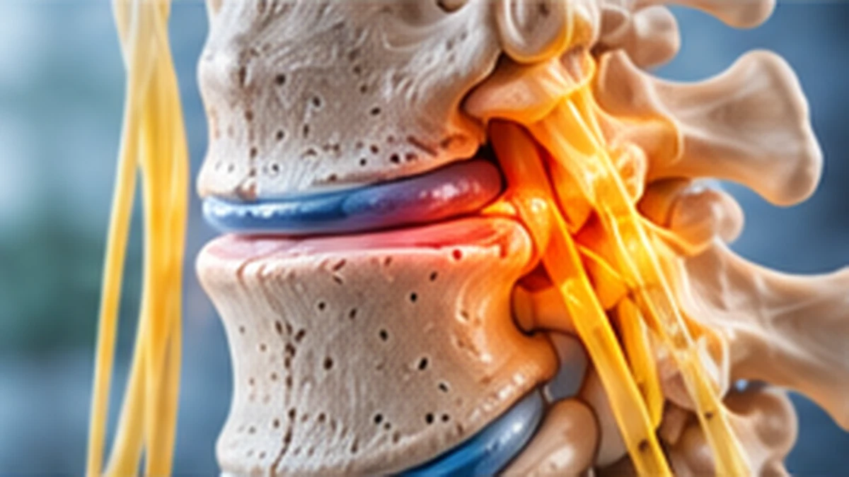
Spinal stenosis means that space within the spinal canal or around a nerve has narrowed. In the lower back, it can contribute to leg pain, heaviness, tingling or walking difficulty, particularly when standing or walking for a period. Narrowing on a scan is not always symptomatic, so treatment depends on function, examination and the pattern of symptoms.
Key points
- Spinal canal narrowing on imaging does not always cause symptoms.
- Walking-related symptoms should be assessed alongside circulation, hip and nerve conditions.
- Treatment can range from activity changes and rehabilitation to selected procedures or surgery.
- New bladder or bowel dysfunction, saddle numbness or rapidly worsening weakness is an emergency.
What is spinal stenosis?
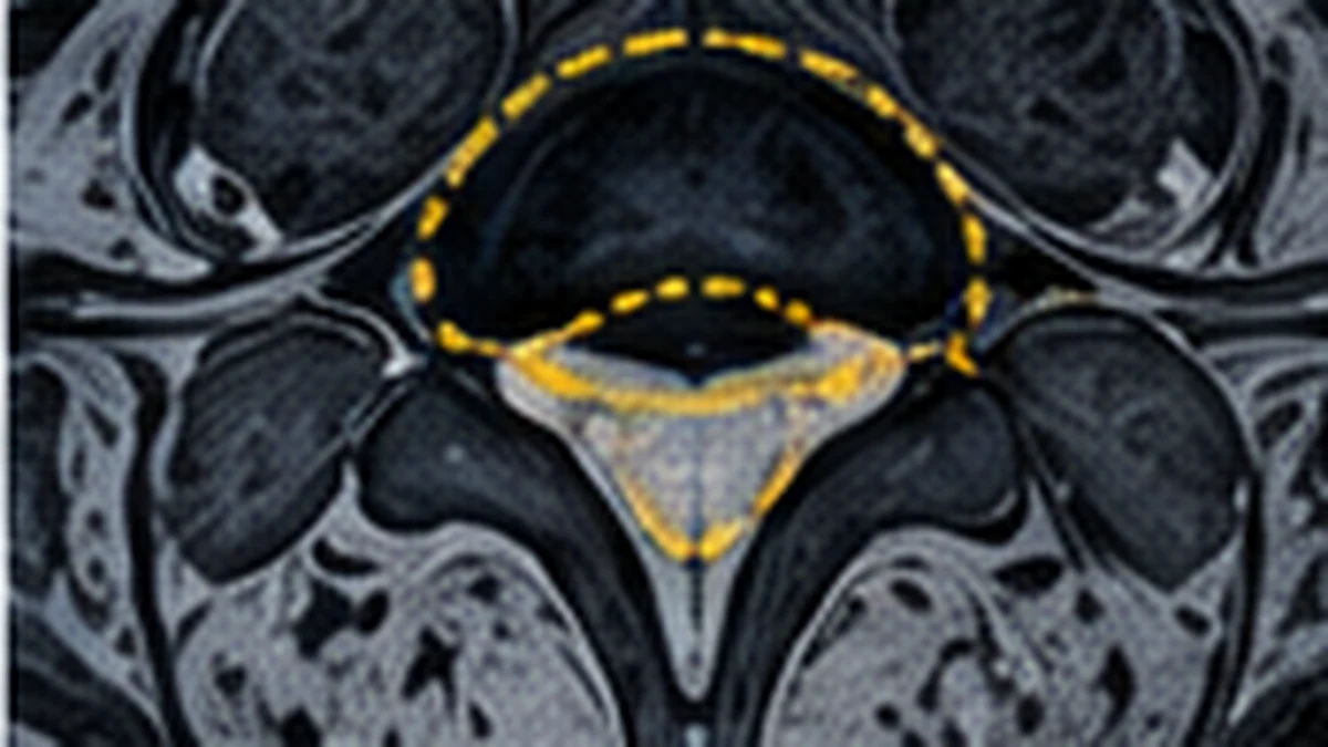
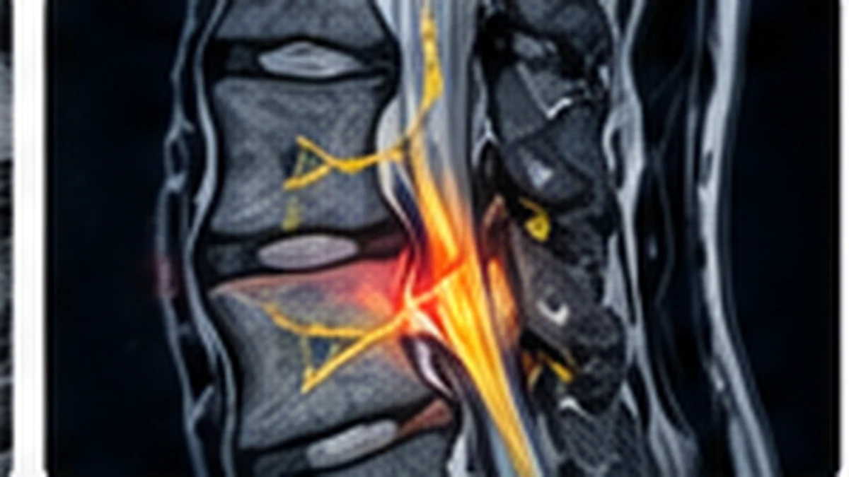
The spinal canal is the space that contains the spinal cord and nerve structures. In the lower back, the cord usually ends higher up and nerve roots travel through the canal. Spinal stenosis describes narrowing of this space or of the side channels where nerves pass. Narrowing may develop as discs lose height, joints enlarge, ligaments thicken or several changes occur together. It is often associated with ageing, but the degree of narrowing does not directly predict how much trouble a person will have. Some people have notable stenosis on MRI and remain active; others have symptoms that significantly limit walking. The location matters: central canal narrowing, narrowing around an exiting nerve and narrowing at several levels can produce different patterns. A clinician needs to connect the scan to the person’s symptoms and examination before deciding whether the finding is clinically important.
Symptoms that may suggest lumbar stenosis
Common symptoms include pain, tingling, numbness, heaviness or weakness in one or both legs when standing or walking. Some people find they need to sit down or lean forward to relieve symptoms. Walking uphill, cycling or bending forward may feel easier for some patients, while standing upright for a long time may be difficult. These patterns can suggest neurogenic claudication, but they are not proof of stenosis. Symptoms may vary from day to day and may affect one leg more than the other. Back pain can occur, but some people mainly notice leg symptoms. Tell the clinician how far you can walk before symptoms start, how long it takes to settle and whether sitting helps. Report persistent numbness, falls or weakness clearly. A sudden change is more concerning than a stable long-term pattern and should prompt reassessment.
Other causes of walking difficulty
Walking difficulty has several possible causes, and identifying the correct one matters because treatment differs. Reduced blood flow in the legs can cause exertional discomfort that settles with rest. Hip or knee arthritis may limit distance because of joint pain. Peripheral neuropathy can cause numbness or balance difficulty, and other neurological conditions can alter gait. More than one problem may be present, especially in older adults. The clinician may ask about smoking, diabetes, cardiovascular disease, joint symptoms and whether discomfort improves with sitting or simply stopping. Examination can include pulses, joint movement, strength, sensation and balance. If symptoms do not fit the MRI finding, further assessment may be needed rather than assuming the spine explains everything. Patients should not feel pressured into spine surgery until the likely source of their limitations has been discussed in a clear and evidence-informed way.
How stenosis is diagnosed
Assessment includes the walking and symptom history, a neurological examination and review of functional limits. The clinician checks strength, sensation, reflexes, gait and other possible causes of symptoms. MRI can show the canal, discs, ligaments and nerve roots, and may help identify the levels where narrowing occurs. X-rays can sometimes provide information about alignment or movement; other tests may be appropriate if circulation or peripheral nerve disease is suspected. The image should answer a clinical question. A scan report listing “moderate” or “severe” narrowing does not by itself tell us whether surgery is necessary. It is important to discuss whether the location and degree of narrowing match the symptoms and examination. Bring previous images if available, because comparison can help avoid unnecessary repeat testing and clarify what has changed.
Non-surgical treatment options
For many people with stable symptoms, non-surgical treatment can be a reasonable starting point. A plan may include education, adapting activities that reliably aggravate symptoms, appropriate medication and physiotherapy. The aim is to improve safe mobility, strength, balance and tolerance for daily activities. Some patients benefit from exercises performed in positions that are more comfortable, but programmes must be individualised. A walking plan can begin with manageable intervals and gradually increase as tolerated. Use a support or mobility aid if recommended to reduce fall risk. Pain medicines require consideration of age, kidney function, stomach history, blood pressure and other medicines. Do not assume that every anti-inflammatory medicine is safe for long-term use. If symptoms remain limiting, review the diagnosis and goals with the clinician rather than repeating an ineffective programme indefinitely. Non-surgical care should have a purpose and a plan for reassessment.
Daily activity and pacing
When walking triggers symptoms, people sometimes stop moving almost entirely. That can lead to reduced conditioning and make everyday tasks even harder. A safer approach is often to pace activity within a manageable range, use planned rests and gradually build capacity under clinical guidance. If leaning forward provides relief, the clinician or therapist may help adapt daily tasks while also considering balance and fall risk. A shopping trip, for example, can be divided into shorter segments rather than treated as one endurance test. Keep track of the distance or time before symptoms begin and how quickly they settle. Stop and seek advice if new weakness, falls or significant neurological changes develop. Do not push through severe or rapidly changing symptoms simply to meet a step target. The objective is sustainable function, not a competition with a fitness tracker.
Injections and symptom management
For selected patients, an injection may be discussed as one component of symptom management. Its usefulness depends on the likely pain source, the type of stenosis and the person’s health. An injection may provide temporary relief for some patients, but it does not widen a narrowed canal permanently. The clinician should explain the intended target, expected benefit, limitations, risks and alternatives. If an injection is proposed, ask how success will be judged and what the plan is if improvement is brief or absent. Repeated procedures without a clear rationale deserve reconsideration. Injections are not a substitute for urgent evaluation when severe neurological warning signs are present. Treatment decisions should also account for medicines that affect bleeding, diabetes and other conditions that may influence procedural safety.
When decompression surgery may be considered
Surgery may be considered when stenosis causes persistent, significant limits on walking or daily life despite appropriate non-surgical care, and clinical findings and imaging identify a matching source. Progressive neurological deficits or emergency symptoms require a different urgency. Decompression aims to create more room for the affected nerve structures. The exact procedure depends on the level, anatomy, stability and whether other spinal problems are present. Some patients may need additional stabilisation, while others do not. Surgery can improve symptoms for appropriately selected patients, but it cannot reverse every age-related change or guarantee a completely pain-free back. The surgeon should explain the intended benefit, possible complications, expected recovery, alternatives and factors that could affect results. A scan showing narrowing is not, by itself, an instruction to operate. The decision should reflect the person’s function, risks and preferences.
Risks and recovery after treatment
Both non-operative and operative approaches have limitations. Rehabilitation may take time and symptoms may fluctuate. Surgery carries risks such as infection, bleeding, dural tear, nerve injury, blood clots, anaesthesia-related complications, persistent symptoms and possible further surgery. The risk profile depends on the specific procedure and the patient’s health. Recovery instructions may include walking, wound care, lifting limits, driving restrictions and follow-up appointments. Do not use another patient’s timeline as a promise for your own recovery. Some patients notice improvement in leg symptoms before endurance returns; others need a longer rehabilitation period. Contact the surgical team for worsening neurological symptoms, wound concerns, fever or other problems listed in discharge instructions. Clear pre-operative counselling should explain what is expected and which changes are not expected.
Symptoms that need urgent care
Seek emergency assessment if you develop new bladder or bowel dysfunction, difficulty passing urine with spinal symptoms, numbness around the groin or saddle area, or rapidly worsening weakness. New severe walking difficulty, repeated falls or sudden loss of coordination also needs prompt medical attention. Fever, recent serious infection, major trauma, a history of cancer or unexplained weight loss may require urgent assessment when associated with spinal pain. These symptoms do not all point to stenosis, but they can indicate a serious condition that should not wait for a routine appointment. If you cannot walk safely, arrange help rather than attempting to travel alone. Explain when the change began and whether it is progressing.
Preparing for a consultation in Navi Mumbai
Before a consultation for spinal stenosis in Kharghar or Navi Mumbai, record how long or how far you can walk, what symptoms appear, whether sitting relieves them and whether balance or strength has changed. Bring MRI reports and images, medicine details and information about diabetes, vascular disease or joint problems. Ask whether the pattern fits spinal stenosis or whether another cause needs investigation. Ask what the initial treatment is intended to improve and when it should be reviewed. If surgery is discussed, ask which level is being treated, whether decompression alone is appropriate and what risks apply to your health. A useful plan should help you understand the next step, the expected measures of progress and warning signs that should prompt earlier review.
Frequently asked questions
No. Some people have narrowing on imaging without symptoms. Diagnosis depends on the symptom pattern, examination and relevant imaging.
Some people with lumbar stenosis find that bending forward reduces symptoms, but this pattern is not diagnostic on its own.
It may improve strength, balance and functional tolerance for selected patients. The programme should be tailored to symptoms and other health conditions.
It may be discussed when symptoms substantially limit function despite suitable care or when neurological findings and imaging support intervention.
New bladder or bowel dysfunction, saddle numbness or rapidly worsening weakness require emergency assessment.
Questions about your spine symptoms?
Arrange a consultation to discuss symptoms, examination findings and available treatment options with the clinic team.
Educational information only; this page does not replace an individual medical assessment. Medical information is provided for general education and does not replace an individual clinical assessment.